
Brain Tumours
Clinical Interests Last updated: Author: Yücel Doğruel, MD, Neurosurgeon
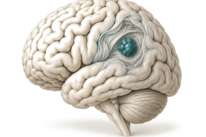
Finding a mass in the brain does not always mean that a malignant tumour is present or that surgery is required. Brain tumours differ from one another in the tissue they arise from, their location, their growth characteristics and their relationship to the surrounding brain structures. Each patient’s treatment is therefore assessed according to his or her own clinical and radiological features.
The principal aim in brain tumour surgery is to establish the diagnosis and treat the disease while preserving neurological function as far as possible. In some patients surgical removal of the tumour is appropriate; in others a biopsy, radiotherapy or follow-up with regular imaging may be the better option.
Which brain tumours?
Gliomas
Tumours arising from the brain tissue itself, such as astrocytoma, oligodendroglioma and glioblastoma. Because their biological features and growth behaviour differ, so do the treatment approaches.
Brain metastases
These develop when cancers of the lung, breast, kidney and other organs spread to the brain. The decision on surgery takes into account the number, size and location of the lesions and the status of the patient’s systemic disease.
Meningiomas
Tumours arising from the membranes covering the brain. Some grow slowly and can be followed up, while surgical treatment may be considered for meningiomas that are growing or causing neurological symptoms.
Deep-seated and intraventricular tumours
Tumours that develop in anatomically demanding regions such as the thalamus, the basal ganglia, the cerebral ventricles and the pineal region. The surrounding vessels, nerve pathways and functional brain structures are important in determining the surgical approach.
Brainstem tumours
The brainstem contains structures related to vital functions such as movement, sensation, swallowing and breathing. In assessing lesions in this region, the type of tumour, its location and its relationship to the surrounding tissue are important in determining the surgical options.
What symptoms can brain tumours cause?
The symptoms of brain tumours vary with their location, their size and their effect on the surrounding tissue. Headache, epileptic seizures, weakness in an arm or leg, disturbances of speech and vision, balance problems and cognitive changes may occur.
These symptoms are not specific to brain tumours. Diagnosis requires neurological examination together with appropriate imaging studies.
New-onset seizures, sudden neurological deficits or changes in consciousness require urgent medical assessment.
When is surgery needed for a brain tumour?
The decision to operate is not based on the size of the tumour alone. The type of tumour, its location, its growth rate, the symptoms it causes, its relationship to neurological function and the patient’s general condition are considered together.
Surgery may have different aims:
- To establish the diagnosis: obtaining tissue so that the pathological and molecular features of the tumour can be determined.
- To remove the tumour: removing the whole tumour, or as much of it as is possible within safe limits, in suitable cases.
- To relieve pressure: reducing the pressure on the brain tissue and the neurological symptoms it causes.
- To prepare for other treatments: providing the diagnostic or surgical groundwork for radiotherapy and systemic treatment where required.
In some tumours a biopsy, radiosurgery or regular MRI follow-up is preferred to surgical removal. There is therefore no single treatment approach for all brain tumours.
How is surgery planned?
Preoperative assessment in brain tumour surgery goes beyond determining the size and location of the tumour. Its relationship to the surrounding vessels, to the functional areas of the cerebral cortex and to the deep white-matter tracts also influences the choice of surgical approach.
Contrast-enhanced MRI of the brain is the principal imaging method. Advanced MRI sequences, tractography, CT and vascular imaging may be used where required.
In tumours close to the speech and motor areas, awake craniotomy and intraoperative neuromonitoring are used according to the features of the case. In anatomically complex lesions, patient-specific three-dimensional models are built in selected cases and the surgical approach is planned on them before the operation. Neuronavigation and intraoperative ultrasonography, in turn, support the localisation of the surgical target and the assessment of the resection during surgery.
The surgical goal is not the removal of the whole tumour in every case. In lesions closely related to speech, movement and other critical brain functions in particular, the balance between the extent of resection and the preservation of neurological function is what matters.
The postoperative period and additional treatment
Pathological and, where required, molecular examination of the tissue obtained at surgery helps to establish the definitive diagnosis and to determine the subsequent treatment options.
Depending on the features of the tumour, radiotherapy, radiosurgery, chemotherapy or other systemic treatments may be needed. In gliomas and brain metastases in particular, the treatment process may call for joint assessment by the relevant specialties.
Postoperative MRI and regular clinical follow-up are important for assessing the result of surgery and for the long-term monitoring of the disease.
Frequently asked questions
Is a brain tumour always cancer?
No. Brain tumours may be benign or malignant. Even a benign tumour, however, may require treatment depending on where it is and how it affects the surrounding tissue.
Can the whole tumour be removed?
This depends on the type of tumour, its location and its relationship to the important brain structures around it. Some tumours can be removed completely, while in some patients a limited resection is preferred in order to preserve neurological function.
Does every brain tumour need surgery?
No. Follow-up with regular imaging, biopsy, radiotherapy or other treatments may be more appropriate for some patients. The treatment decision requires individual assessment.
Will other treatments be needed after surgery?
Additional treatment may be needed depending on the pathological and molecular features of the tumour, the result of surgery and the patient's general condition. Each patient's follow-up and treatment programme is different.
Information needed for the first assessment
It is important that the current brain MRI and any previous studies can be reviewed, if possible together with the digital image files (DICOM). If surgery, radiotherapy or oncological treatment has been given before, the relevant reports also help in understanding the treatment history.
For referring physicians
A DICOM copy of the contrast-enhanced MRI, perfusion and spectroscopy studies where available, the current stage of the systemic disease and the oncology note in cases of metastasis, details of previous surgery and radiotherapy, and the pathology report make the assessment easier. Professional correspondence: yucel@yuceldogruel.com
Related publications
The information on this page is for general purposes only. Diagnosis and treatment decisions are made on the basis of each patient’s individual assessment.