
Aneurysms and Cerebrovascular Disease
Clinical Interests Last updated: Author: Yücel Doğruel, MD, Neurosurgeon
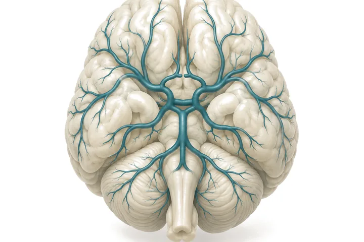
Cerebrovascular diseases are conditions that affect the structure of the vessels supplying the brain or the circulation of blood through them, and that in some cases can lead to brain haemorrhage or loss of neurological function. Brain aneurysms, arteriovenous malformations (AVMs) and cavernomas belong to this group.
Not every cerebrovascular disease requires surgical intervention. The treatment decision weighs the type of disease, the vascular anatomy, any history of haemorrhage, the patient’s age and general health, and the risks of the treatments that could be applied.
Which diseases?
Brain aneurysms
Balloon-like dilatations of a cerebral artery that form where a weak area of the vessel wall expands. Some aneurysms are found without having caused any symptoms, while others may grow or rupture and cause a subarachnoid haemorrhage.
In unruptured aneurysms, regular follow-up, microsurgical clipping or endovascular treatment from within the vessel may be considered. The choice of treatment depends on the features of the aneurysm and the patient’s risk factors.
Arteriovenous malformations (AVMs)
Abnormal connections between the arteries and veins of the brain that form without the normal capillary network in between. They may present with brain haemorrhage, epileptic seizures or neurological symptoms, or be found on imaging performed for another reason.
Treatment options include microsurgical removal, endovascular embolisation, stereotactic radiosurgery or a combination of these. In some patients observation is preferred to intervention.
Cavernous malformations (cavernomas)
Lesions made up of clusters of abnormal small vessels that can occur in the brain or the brainstem. They may cause haemorrhage, epileptic seizures or neurological symptoms.
The treatment decision depends on the location of the cavernoma, previous haemorrhages and the patient’s neurological condition. In brainstem cavernomas in particular, the potential benefits and the neurological risks of surgery are weighed with great care.
When does a brain aneurysm need treatment?
Finding an aneurysm does not necessarily mean that it must be operated on. In unruptured aneurysms in particular, the essential decision is a balance between the likelihood of future haemorrhage and the risks of intervention.
In assessing the risk of haemorrhage from an unruptured aneurysm, its size, location and shape, any growth over time, the patient’s age, family history, hypertension and smoking are taken into account. The configuration of the aneurysm neck and its relationship to the surrounding vessels are important in choosing the appropriate treatment.
In unruptured aneurysms, follow-up with regular imaging may be appropriate. Where treatment is required, microsurgical or endovascular methods are considered.
In ruptured aneurysms, because of the risk of re-bleeding, urgent assessment and appropriate treatment at the earliest possible stage are essential.
In the event of a sudden, very severe headache, loss of consciousness, new weakness or disturbance of speech, emergency medical help should be sought by calling 112 without waiting for an outpatient appointment.
Diagnosis and surgical planning
CT angiography, MR angiography and digital subtraction angiography (DSA) are used in assessing cerebrovascular disease. CT and MR angiography image the cerebral vessels without placing a catheter in the artery. DSA is a catheter-based study that allows a more detailed assessment of the vascular anatomy and blood flow. Which method is needed depends on the patient’s clinical condition and the imaging already available.
Diagnostic cerebral angiography provides important information for deciding the treatment strategy, particularly in complex aneurysms and AVMs. It is not needed in every patient; the decision is made on the basis of the existing images and the clinical requirements. Where required, this study is performed and interpreted by the physician who plans the surgery, so that the findings on vascular anatomy are carried directly into the treatment plan.
In surgical planning, the neck of the aneurysm, its relationship to the surrounding vessels, the course of the vessels and the possible surgical corridors are assessed in detail. In selected cases, patient-specific three-dimensional models are built from CT angiography and DSA data so that the position of the craniotomy, the direction of the surgical approach and the clip placement options can be examined before the operation.
Surgical treatments
Microsurgical aneurysm clipping
In aneurysm clipping, a purpose-made clip is placed across the neck of the aneurysm under the microscope so as to exclude the aneurysm from the circulation. Preserving the vessels around the aneurysm and the small branches arising from them is essential during surgery.
Where required, vessel patency and blood flow can be assessed with micro-Doppler and indocyanine green (ICG) video angiography.
Endovascular treatments
In endovascular methods the aneurysm is reached from within the vessel with catheters. Depending on the structure of the aneurysm, coils, stent-assisted techniques or flow-diverting devices may be used.
Microsurgical clipping and endovascular methods are weighed according to the anatomical features of the aneurysm and the patient’s clinical condition. Some aneurysms can be treated by either method, while for others one particular method may be more suitable.
AVM surgery
The aim in AVM surgery is to remove the abnormal vascular tangle without damaging the normal brain circulation around it. Suitability for surgery is assessed with regard to the size, location and venous drainage of the malformation and its relationship to functional brain areas.
Cavernoma surgery
In cavernoma surgery, the location of the lesion and whether it has bled before are important. In brainstem and deep-seated cavernomas, microsurgical neuroanatomy is of particular importance in determining the entry point and the approach.
Cerebral bypass surgery
Microsurgical vascular connections intended to re-establish or preserve blood flow in the cerebral vessels. They may be considered in some complex aneurysms and in specifically selected vascular diseases. Bypass is not a routine treatment applied to every vascular occlusion.
Follow-up after treatment
Follow-up after treatment is planned according to the type of disease and the method used.
After aneurysm treatment, vascular imaging is used to confirm that the aneurysm has been excluded from the circulation and to assess any other vascular structures that need to be checked. In AVMs and cavernomas, follow-up consists of neurological examination and appropriate imaging according to the features of the disease.
Frequently asked questions
Does every brain aneurysm need surgery?
No. For some aneurysms, observation with regular imaging is appropriate. The treatment decision is made by weighing the risk of haemorrhage from the aneurysm against the possible risks of intervention.
Is clipping or endovascular treatment more suitable?
There is no single method that applies to every patient. The neck, size and location of the aneurysm, its relationship to the surrounding vessels and the patient's characteristics determine the choice of treatment.
Does a brain aneurysm cause headache?
Most aneurysms cause no symptoms and are found on studies performed for other reasons. A headache on its own does not mean that an aneurysm is going to bleed. A sudden, very severe headache, however, may be a sign of brain haemorrhage and requires urgent assessment.
Are AVM and cavernoma the same disease?
No. Although both are vascular malformations, their vascular structure, imaging features, haemorrhage risk and treatment approaches differ.
Can cerebrovascular disease be treated without surgery?
Yes. Depending on the type of disease, options such as follow-up with imaging, endovascular treatment or radiosurgery may be considered. Each patient's treatment decision is made individually.
Information needed for the first assessment
It is important that the current CT angiography, MR angiography or DSA images can be reviewed, in DICOM format if possible, together with any previous studies. If there is a history of brain haemorrhage, surgery or radiosurgery, the relevant reports also help in understanding the treatment history.
For referring physicians
In aneurysm cases, sharing the CT angiography or MR angiography images in DICOM format makes the first assessment easier; it is not necessary for every patient to have undergone DSA beforehand. In AVM cases the existing angiographic studies are important, and in cavernomas images that include the appropriate MRI sequences. Professional correspondence: yucel@yuceldogruel.com
Related publications
The information on this page is for general purposes only. Diagnosis and treatment decisions are made on the basis of each patient’s individual assessment.