
Surgical Neuroanatomy
Research and Teaching Last updated:
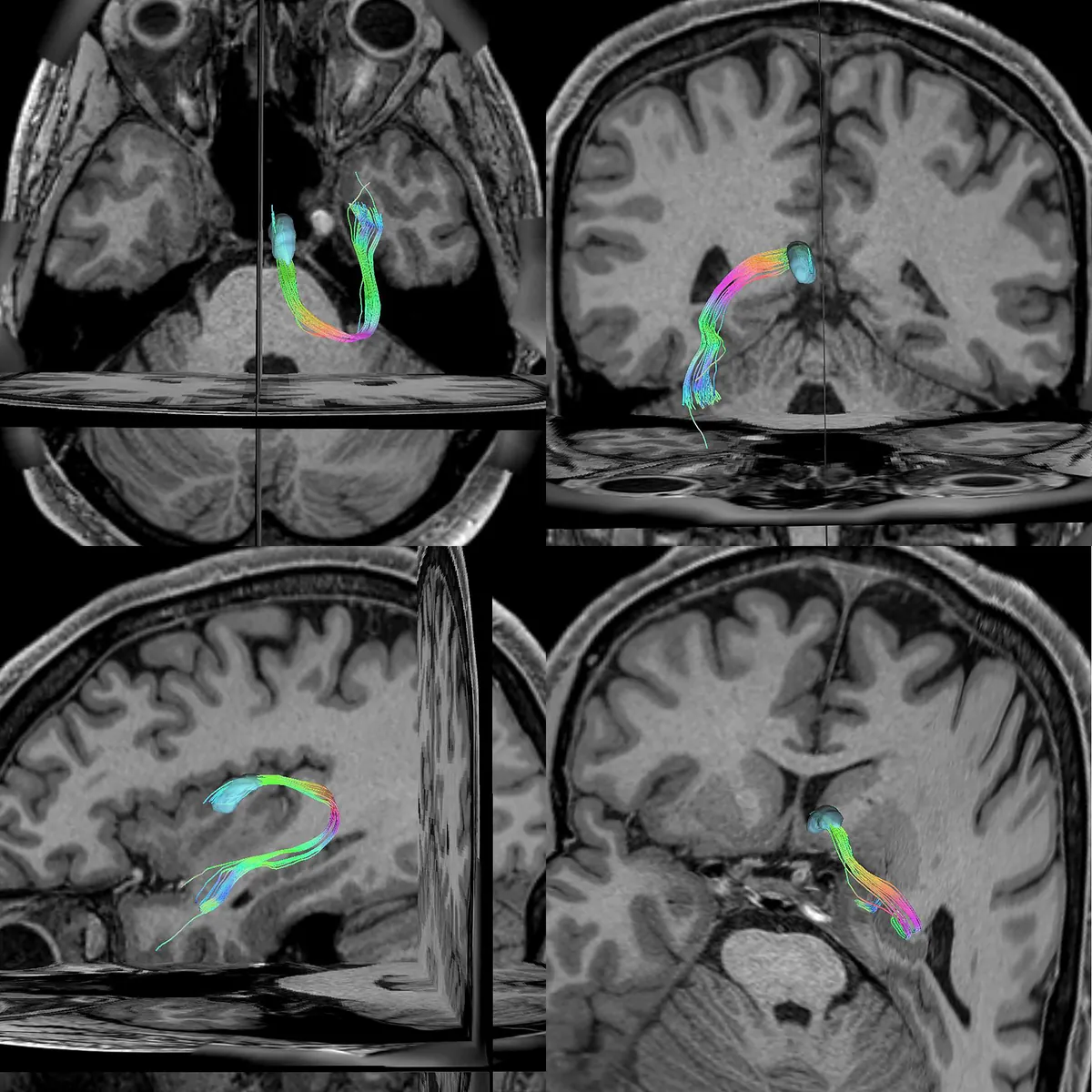
Surgical neuroanatomy is the field that studies the relationships between the vascular, neural and white-matter structures of the brain and their significance for surgical approaches. In deep-seated brain lesions and skull base surgery in particular, a detailed understanding of the anatomical corridors leading to the target region is essential.
Studies in microsurgical neuroanatomy and white-matter dissection make it possible to investigate the anatomical basis of surgical approaches and to apply this knowledge in surgical planning.
Laboratory and research work
As part of his training and research in microsurgical neuroanatomy, he has carried out studies on white-matter dissection, cranial nerve anatomy, cerebrovascular anatomy and skull base approaches in the laboratories of the University of Athens, Acıbadem University and Yeditepe University.
In the joint research programme of The Neurosurgical Atlas and Yeditepe University, he took part in research on surgical neuroanatomy, anatomical visualisation and the preparation of surgical teaching material.
Details of these training and research programmes are given on the About page.
Principal research areas
White-matter anatomy and fibre dissection
The anatomical organisation of the white-matter tracts connecting the different regions of the brain is important in planning surgical approaches. The course of these tracts, their connections and their relationship to surgical corridors are investigated by cadaveric dissection and MR tractography.
Cerebrovascular anatomy
The microsurgical anatomy of the arterial and venous circulation of the brain is one of the fundamental areas of study for surgical approaches to aneurysms, vascular malformations and deep-seated lesions in particular.
The research includes surgical techniques for preserving the cerebellar tentorial bridging veins and studies of the deep cerebral venous anatomy.
Deep brain and skull base approaches
The anatomical features of the surgical corridors to the thalamus, the brainstem, the ventricles, the pineal region and the skull base are studied. In choosing the surgical approach, the relationships between the nerve pathways, the vessels and the surrounding functional structures are assessed together.
Three-dimensional visualisation of microsurgical anatomy
Converting the anatomical information obtained from cadaveric dissections and radiological data into three-dimensional models is a further area of work directed at teaching surgical neuroanatomy.
This research is also covered on the 3D Modelling and Virtual Reality page.
Relevance to surgical planning
Surgical neuroanatomy studies contribute to the assessment of the anatomical limits, advantages and constraints of the different surgical approaches.
The anatomical knowledge gained in the laboratory is interpreted together with the patient’s MRI, CT and vascular imaging findings and used in planning the surgical approach.
The study on the tentorial bridging veins appeared on the cover of the January 2024 issue of the Journal of Neurosurgery, and the angular gyrus study on the cover of the January 2023 issue of Brain Structure and Function.